
Quick Actions
Contents
Neuron Structure & Function
This topic introduces the structure and functional organization of neurons, the fundamental signaling units of the nervous system. It explains how neurons receive, integrate, conduct, and transmit information through electrical and chemical processes. Students will learn the structural components of a neuron, how action potentials are generated and propagated, and how neurons communicate at synapses. This note provides a strong scientific foundation for understanding neural physiology, neurobiology, and clinical disorders affecting nerve function.
Learning Objectives
- Describe the major structural components of a neuron.
- Explain how resting membrane potential is established and maintained.
- Discuss the phases of an action potential and the ionic changes involved.
- Differentiate between graded potentials and action potentials.
- Describe synaptic transmission and the roles of neurotransmitters.
- Explain myelination and how it influences conduction velocity.
Key Points to Remember
- Neurons are specialized cells that transmit electrical and chemical signals.
- Dendrites receive input while axons conduct impulses away from the cell body.
- Resting membrane potential is maintained by ion gradients and selective permeability.
- Action potentials follow an all or none principle.
- Synaptic transmission involves release of neurotransmitters at synaptic terminals.
- Myelination increases the speed of impulse conduction.
Introduction to Neuron Structure and Function
Neurons are highly specialized excitable cells that form the communication network of the nervous system. Their unique structural organization allows them to receive signals, generate impulses, and transmit information efficiently over short or long distances.
Each neuron is composed of distinct regions that contribute to its overall function, and understanding these structures is essential for studying neural physiology and human nervous system function.
Structural Components of a Neuron
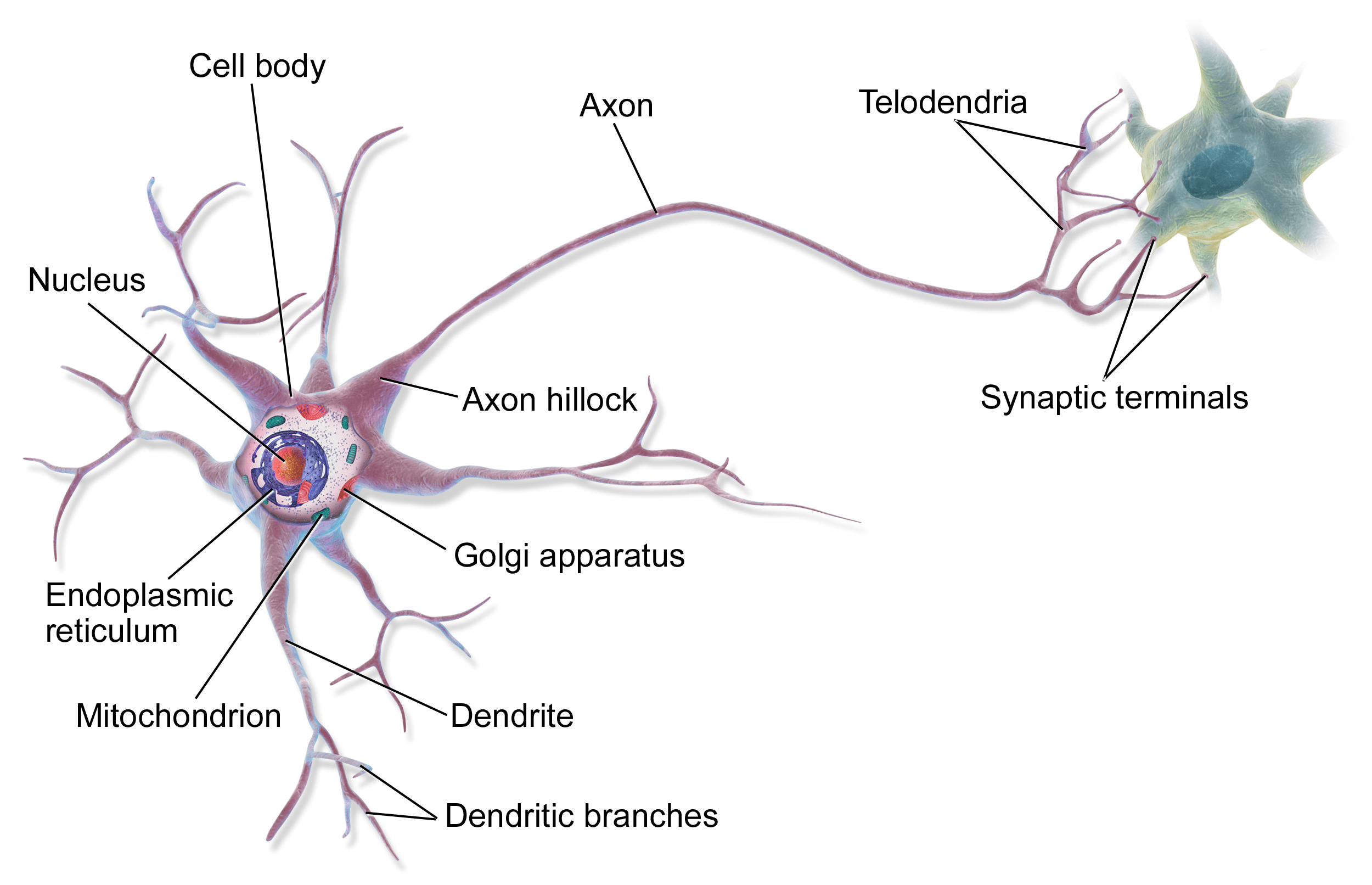
1. Cell Body
The cell body, also called the soma, contains the nucleus and most of the cytoplasmic organelles. It integrates incoming signals from the dendrites and plays a central role in protein synthesis. Nissl bodies, which are abundant rough endoplasmic reticulum fragments, are prominent in the soma and are responsible for synthesis of neurotransmitter molecules and structural proteins.
2. Dendrites
Dendrites are branching cytoplasmic projections that increase the receptive surface area of the neuron. They receive chemical signals from other neurons through synapses and convert them into graded potentials. These signals travel toward the cell body where they are processed. The number and complexity of dendrites vary depending on the type of neuron and its functional role.
3. Axon
The axon is a long process that conducts impulses away from the cell body. It originates from a region called the axon hillock, which is the site where action potentials are initiated when threshold is reached. The axon may branch to form collaterals and terminates in synaptic boutons, which release neurotransmitters.
A helpful mnemonic for remembering the functional flow in a typical neuron is “D S A T” which stands for Dendrite receives, Soma integrates, Axon conducts, Terminal transmits.
4. Myelin Sheath
Some axons are wrapped by myelin, a lipid rich insulating covering formed by Schwann cells in the peripheral nervous system and oligodendrocytes in the central nervous system. Myelin increases the velocity of impulse conduction by allowing current to leap between nodes of Ranvier in a process called saltatory conduction. Myelinated fibers conduct impulses more rapidly than unmyelinated ones, making myelination essential for fast reflexes and coordinated movement.
5. Synaptic Terminals
Synaptic terminals form junctions with other neurons, muscles, or glands. They contain synaptic vesicles filled with neurotransmitters. When an action potential reaches the terminal, it triggers release of neurotransmitters into the synaptic cleft, enabling communication with target cells.
Resting Membrane Potential
Definition and Importance
The resting membrane potential is the electrical potential difference across the neuron's membrane when it is not transmitting a signal. It is usually about -70 millivolts, meaning the interior of the neuron is more negative than the exterior. This electrical potential sets the stage for generation of action potentials.
Factors Responsible for Resting Potential
Selective membrane permeability
Potassium ions move more freely across the membrane than sodium ions because there are more potassium leak channels. As potassium diffuses out, it leaves behind negatively charged proteins, contributing to the negative internal charge.
Sodium potassium pump
The sodium potassium pump continuously moves three sodium ions out and two potassium ions in. This maintains ionic gradients and helps sustain the negative resting potential.
A simple mnemonic is “3 out, 2 in makes the inside thin” to remember that more positive charges leave the cell than enter it.
Graded Potentials
Graded potentials are small changes in membrane potential that vary in size depending on stimulus strength. They occur mainly in dendrites and the soma. They are not all or none and do not propagate long distances. Instead, they decay over space and time. Graded potentials determine whether the axon hillock will reach threshold to initiate an action potential.
Action Potential
Overview
An action potential is a rapid and brief reversal of membrane potential that travels along the axon. It allows long distance communication within the nervous system. It obeys the all or none principle, meaning it either occurs fully or not at all.
Phases of the Action Potential
1. Depolarization
During depolarization, voltage gated sodium channels open causing sodium to rush into the cell. The membrane potential becomes less negative and eventually positive.
2. Repolarization
As the membrane depolarizes, sodium channels become inactivated and voltage gated potassium channels open. Potassium flows out of the cell restoring the negative membrane potential.
3. Hyperpolarization
Potassium channels remain open slightly longer, causing the membrane potential to become more negative than the resting value before stabilizing.
Refractory Periods
Absolute refractory period
During this time no stimulus can trigger another action potential because sodium channels are inactivated.
Relative refractory period
A stronger than usual stimulus is required to initiate another action potential because the membrane is still hyperpolarized.
A mnemonic often used is “Depo In, Repo Out” referring to sodium ions entering during depolarization and potassium ions exiting during repolarization.
Propagation of Action Potentials
Continuous Conduction
In unmyelinated axons, each segment of the membrane must depolarize sequentially. This process is slower and occurs along the entire length of the axon.
Saltatory Conduction
In myelinated fibers, action potentials jump between nodes of Ranvier where voltage gated channels are concentrated. This increases conduction speed significantly. Saltatory conduction is highly energy efficient and critical for rapid neural signaling.
Synaptic Transmission
Types of Synapses
Chemical synapses
These involve release of neurotransmitters from synaptic vesicles. They are the most common type. Neurotransmitters diffuse across the synaptic cleft and bind to receptors on the postsynaptic membrane.
Electrical synapses
These involve direct transfer of ions through gap junctions. They allow rapid and synchronized communication but are less common in humans.
Events in Chemical Synaptic Transmission
- Arrival of an action potential at the terminal.
- Opening of voltage gated calcium channels.
- Calcium entry triggers fusion of synaptic vesicles with the membrane.
- Neurotransmitter is released into the cleft.
- Neurotransmitter binds to receptors on the postsynaptic membrane.
- Response occurs, either excitation or inhibition.
- Neurotransmitter is removed by reuptake, enzymatic breakdown, or diffusion.
Neurotransmitters
Common neurotransmitters include acetylcholine, glutamate, gamma aminobutyric acid, glycine, dopamine, serotonin, and norepinephrine. Each neurotransmitter has specific receptors and physiological roles.
Myelination and Conduction Velocity
Myelin Formation
Schwann cells wrap around peripheral axons while oligodendrocytes myelinate central axons. Each Schwann cell forms a single segment of myelin while oligodendrocytes can myelinate multiple axons. The myelin sheath is composed mainly of lipids which provide electrical insulation.
Functions of Myelin
Clinical Relevance
Diseases such as multiple sclerosis and Guillain Barré syndrome affect myelin and result in slowed transmission, muscle weakness, and sensory disturbances.
Test Your Knowledge
Complete the quiz to check your understanding.
Ready to take the quiz?
Please log in to save your progress and see how you stack up!
Log In to ContinueAdditional Resources
See More Resourses
Watch more on Youtube